Why Was the MRI Invented?
Join me in discovering the fascinating story of how the MRI was invented and revolutionized medicine!
Source www.starimagingindia.com
The Invention of the MRI: A Revolution in Medical Diagnosis
The Magnetic Resonance Imaging (MRI) has revolutionized the field of medical diagnosis by allowing doctors to take detailed images of the human body without using harmful x-rays. It is a non-invasive technology that enables the diagnosis of a wide range of medical conditions, from cancer to neurological disorders.
Overview of the MRI
The MRI is a medical imaging technique that uses a strong magnetic field and radio waves to generate images of organs and tissues inside the body. The patient lies inside a machine that contains a powerful magnet, which causes the protons in the body's atoms to align with the magnetic field. Radio waves are then sent through the body, causing the protons to produce a signal that is interpreted by a computer to create images of the body's internal structures.
Discovery of Nuclear Magnetic Resonance (NMR)
The development of the MRI was based on the discovery of Nuclear Magnetic Resonance (NMR), a technology that was first discovered in the 1940s by two physicists, Felix Bloch and Edward Mills Purcell. They received the Nobel Prize in Physics in 1952 for their groundbreaking discovery, which allowed scientists to study the structure of materials at the atomic level.
Birth of the MRI
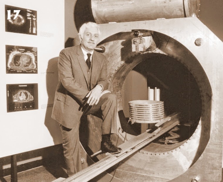
The MRI was invented by Dr. Raymond Damadian, a physicist and medical doctor at the State University of New York. He filed a patent for his invention in 1972, after realizing that the NMR could be used to create images of the human body. His research focused on using the NMR to detect cancerous tissue in the body, as he believed that cancer cells would have different magnetic properties to healthy cells.
After almost a decade of research and testing, Damadian and his team performed the first human MRI scan in 1977 in the United States. It took over five hours to generate an image of a human body, but it proved that the technology was viable for clinical use. The first commercial MRI machine was launched in 1980, and since then, the technology has become an essential tool for medical diagnosis and research.
In conclusion, the invention of the MRI has revolutionized the field of medical diagnosis and has saved countless lives around the world. The MRI has enabled doctors to detect medical conditions that were previously undetectable, and it has had a significant impact on medical research. It is a testament to the power of scientific discovery and innovation, and it is an inspiration to the next generation of scientists and inventors.
When Was the MRI Invented?
The invention of the MRI has revolutionized medical diagnostics by allowing doctors to see inside the human body with greater accuracy and detail than ever before.
The MRI technology was first developed in the late 1960s by two scientists, Paul Lauterbur and Sir Peter Mansfield. Their groundbreaking work earned them the Nobel Prize in Physiology or Medicine in 2003.
Paul Lauterbur’s Contribution
Paul Lauterbur was an American chemist who developed the concept of using magnetic fields to generate images of the body. In 1971, he published a paper in the journal Nature, titled “Image Formation by Induced Local Interactions: Examples Employing Nuclear Magnetic Resonance.”
In this paper, Lauterbur proposed a method for creating two-dimensional images of objects by detecting differences in the magnetic properties of their atomic nuclei. This method, which he called “Zeugmatography,” was a precursor to the modern MRI.
Sir Peter Mansfield’s Contribution
Sir Peter Mansfield was a British physicist who constructed the first working MRI scanner in the early 1980s. His work improved upon Lauterbur’s methods by using gradient magnetic fields to encode spatial information and faster imaging techniques.
Mansfield also developed techniques for obtaining functional MRI (fMRI) images, which can show changes in brain activity over time. This method has proved invaluable in studying brain disorders such as Alzheimer’s disease and Parkinson’s disease.
How Does the MRI Work?
The MRI machine uses a combination of strong magnetic fields and radio waves to produce detailed images of internal body structures, such as the brain, spine, and joints.
Principle of the MRI
When a patient enters the MRI machine, they lie down on a table that moves into the machine. The magnetic field of the machine causes the body's hydrogen atoms to align, and then radio waves are sent into the body to disrupt this alignment and create a signal that is captured by the machine's sensors.
These signals are then processed by a computer to create detailed images of the body’s tissues and organs. Different types of tissues produce different signals, allowing doctors to distinguish between bones, muscles, and internal organs.
Advantages and Limitations of MRI
The MRI has several advantages over other imaging technologies, such as it does not use ionizing radiation, it is non-invasive, and can detect subtle abnormalities that other imaging technologies cannot.
However, the MRI also has some limitations, such as it can be relatively expensive, and patients with certain types of metal implants or pacemakers cannot safely undergo MRI procedures.
The MRI has become an essential tool for medical diagnostics, and its development and refinement continue to improve patient care and outcomes.
Did you know about the history of tractors?When Was the MRI Invented?
The MRI, short for Magnetic Resonance Imaging, is a diagnostic medical imaging technique that uses a combination of strong magnetic fields and radio waves to create detailed images of the body's internal structures. It was first developed in the early 1970s by two independent teams of researchers in the United States and the United Kingdom.
Dr. Raymond Damadian, an American physician and medical researcher, is widely credited with the invention of the MRI. In 1970, Dr. Damadian discovered that the NMR (Nuclear Magnetic Resonance) signal differed between normal and cancerous tissue. He used this discovery to develop the first MRI scanner, which he called the "Indomitable".
In 1974, a British researcher named Peter Mansfield developed a new technique for generating MRI images called echo-planar imaging (EPI). This new technique allowed for faster imaging and led to the development of functional MRI (fMRI), which is used to study brain function.
Together, the work of Dr. Damadian and Peter Mansfield paved the way for the development of the modern MRI machines used today.
Applications of the MRI
Diagnostic Applications
The MRI is widely used in medical diagnosis, particularly in the fields of neurology, oncology, and orthopedics. Its ability to produce detailed images of soft tissues and organs makes it a valuable tool in detecting a wide range of conditions, including brain and spinal cord tumors, multiple sclerosis, ligament and muscle tears, and joint disorders such as arthritis.
One of the main advantages of MRI over other imaging techniques like X-rays and CT scans is its ability to produce high-quality images without using ionizing radiation, which can be harmful to patients. This makes it especially useful for imaging pregnant women, children, and other populations who may be more sensitive to radiation exposure.
Research Applications
In addition to its diagnostic applications, the MRI is also used extensively in medical research, particularly in neuroscience and psychology, to study brain structure and function, as well as in studies of the musculoskeletal system.
Researchers use advanced MRI techniques like fMRI to study how the brain responds to different stimuli and to map the neural connections that underlie human behavior and cognition. MRI is also used to study the effects of various drugs and therapies on the brain, as well as to investigate the mechanisms of different diseases and disorders.
Future Developments
Research is ongoing in the field of MRI, with new techniques being developed to improve image quality and patient comfort, as well as to expand its range of applications. One promising area of research is the use of MRI-guided interventions, which allow for more precise targeting during surgeries and other medical procedures.
Another area of research involves the development of superconducting MRI machines, which use superconducting coils to produce stronger magnetic fields and therefore higher-quality images. These machines are still in the experimental phase, but they could have significant implications for the future of MRI technology.
Overall, the wide range of applications and ongoing research in the field of MRI make it an exciting and rapidly evolving area of medicine and medical technology.
Keys: Who invented them and why they are importantThe Impact of the MRI on Medicine
The invention of the Magnetic Resonance Imaging (MRI) machine has revolutionized the medical field, allowing doctors to visualize internal body structures like never before. It has had a significant impact on medical diagnosis and created advancements in the practice of medicine. The MRI has enabled the imaging of living tissues and organs, aiding in the understanding of biological systems. Furthermore, its use has also extended beyond medicine to forensic investigations and the study of cultural artifacts.
Revolutionizing Medical Diagnosis
The MRI has transformed the way doctors diagnose several conditions, especially those affecting the brain and spinal cord. Previously, doctors had to rely on X-rays, computed tomography (CT) scans, and other imaging methods that weren't as efficient in detecting certain conditions. With the MRI, doctors can see in detail the structures inside the body, including soft tissues such as muscles and nerves. The increased accuracy in diagnosis has helped in the detection of a wide range of conditions such as tumors, stroke, and multiple sclerosis.
Advancing the Practice of Medicine
The MRI not only improves diagnosis but has also created advancements in the practice of medicine. The machine's ability to create detailed images of the human body's inner workings has led to the development of new surgical techniques and more targeted therapies. Physicians can now detect diseases earlier, which enhances their effectiveness in treating certain conditions. For example, the use of MRI-guided biopsy has enabled early diagnosis of breast cancer and other tumor growths. In addition, the MRI has aided in understanding biological systems and created avenues for further research, allowing doctors to better understand how the body works and how diseases develop.
Broader Societal Implications
The MRI's impact is not just limited to the medical field. Its non-invasive and high-resolution imaging abilities have made it useful in other areas such as the investigation of crime and study of cultural artifacts. In forensic investigations, the MRI is used to detect hidden evidence such as bone fractures and foreign objects. Additionally, the MRI is utilized in the study of cultural artifacts such as ancient paintings to detect their authenticity and condition without damage to the original work.
In conclusion, the invention of the MRI machine has had a tremendous impact on the medical field and beyond. Its creation has revolutionized medical diagnosis, advanced the practice of medicine, and extended its research capabilities to broader societal applications. MRI machines have become some of the most critical diagnostic and research tools of our time, and their continued advancement will further aid in medical progress and innovation.
Invention of MRI and video recording has much in common!