Where Was MRI Invented?
Discover the Birthplace of MRI: A Fascinating Journey into the History of Medical Imaging
Source www.longislandpress.com
Where Was the MRI Invented?
The MRI Definition
Magnetic Resonance Imaging, or MRI, is a diagnostic test that uses a magnetic field and radio waves to create detailed images of internal body structures. The images produced by MRI can help healthcare professionals diagnose and treat a wide range of medical conditions, from cancer and heart disease to brain injuries and joint problems.
The History of MRI
The initial concepts behind MRI were first proposed in the early 20th century by physicists Isidor Rabi and Felix Bloch. However, it wasn't until the 1970s that the technology for producing MRI images started to come together.
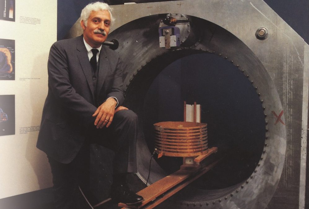
In 1971, American physicist Raymond Damadian developed the first MRI scanner, which he dubbed the "Indomitable." Damadian's early work on MRI involved using nuclear magnetic resonance (NMR) to study changes in the body's chemistry in response to disease. He discovered that cancerous tissue displayed a unique NMR signal, which would eventually form the basis for MRI's diagnostic capabilities.
By the mid-1970s, Damadian had built a prototype of his MRI scanner that was capable of producing rudimentary images of the human body. However, it wasn't until 1977 that he achieved his first successful full-body MRI scan on a human subject. This breakthrough led to the development of MRI as a useful diagnostic tool, and it quickly became a popular alternative to X-rays and other imaging techniques.
The Location of the First MRI Scanner
Contrary to popular belief, Raymond Damadian did not invent the MRI scanner in a high-tech laboratory with state-of-the-art equipment. Instead, he built his first working MRI scanner in a makeshift laboratory in his Brooklyn basement.
With no funding and little support from the scientific community, Damadian spent years tinkering with NMR machines and building his own equipment from spare parts. His early experiments often involved using himself as a test subject, as he struggled to convince others of the potential of his invention. However, his persistence eventually paid off.
The first successful human MRI scan was performed using a prototype of Damadian's scanner at Downstate Medical Center in Brooklyn, New York. The patient was a young man with a suspected bone tumor, and the scan revealed a mass on his hip that was later confirmed to be cancerous.
Today, MRI technology is widely used in hospitals and clinics around the world, and it has revolutionized the field of diagnostic medicine. In 1984, Damadian's company, Fonar Corporation, became the first to commercialize MRI technology for diagnostic use, and the technology has continued to evolve and improve over the years.
Where Was The MRI Invented?
The MRI or Magnetic Resonance Imaging was invented in multiple locations and by multiple scientists. In 1971, Raymond Damadian discovered that cancerous and healthy tissues had different levels of relaxation time on the nuclear magnetic resonance. He performed the experiment on a live mouse, comparing the NMR signal from different organs in the mouse. The result of the experiment showed that different tissues give off distinct NMR signals. He proposed that the nuclei in tumors would have continuous signal and that this could be used to diagnose cancer.
In the same year, Paul Lauterbur proposed a method to produce cross-sectional images using NMR. He proposed that one could scan the object in slices, then combine the data from each slice to build a three-dimensional representation of the object. His concept illustrated that one can manipulate the information contained in the NMR signal by applying different magnetic fields at different locations.
Later, Peter Mansfield discovered that one can encode spatial location information into an NMR signal by incorporating magnetic gradients into the magnetic field. Through his finding, he developed echo-planar imaging, an approach to quickly constructing an image using many gradients. This is commonly used in MRI machines to date.
How Do MRI Scanners Work?
The Basic Principles of MRI
As previously mentioned, the human body is made up largely of water molecules that contain hydrogen atoms. When a magnetic field is introduced, hydrogen nuclei align themselves based on the strength of the magnetic field. The stronger the magnetic field, the better-aligned the hydrogen nuclei are. Radio waves are then introduced to the body at a specific frequency and directed towards the hydrogen atoms. This causes the hydrogen atoms to resonate, emitting a signal that is detected by the MRI scanner. The time it takes for the hydrogen atoms to realign themselves determines the strength of the signal. The scanner then uses mathematical algorithms to convert this information into an image.
The Different Types of MRI Scanners
MRI scanners vary in size, strength, and shape. The most common types include the closed-bore systems, open MRI machines, and upright MRI machines. The closed-bore MRI machines are the most common type and are the ones that most people think of when they imagine an MRI. The patient lies down on a table that slides into the tunnel, where the machine is located. Open MRI machines have an open design, and the patient is not entirely enclosed, making the patient feel more comfortable. Upright MRI machines are not as common and are used for specific types of imaging, such as spinal issues and the musculoskeletal system.
The Advantages and Disadvantages of MRI Imaging
MRI imaging is a non-invasive and painless procedure that produces detailed images of soft tissues and organs. It has become an essential tool in diagnosing a wide range of medical conditions. However, the disadvantages of MRI imaging may outweigh the advantages in certain situations. MRI machines are costly, and the procedure is time-consuming compared to X-ray and CT scans. People with certain medical conditions like implanted devices or heart pacemakers cannot undergo an MRI scan due to the magnetic fields involved in the process.
In conclusion, the MRI machine has revolutionized medical imaging by providing incredible insight into the human body without making any incisions. The invention of the MRI is a result of years of research from scientists who dedicated their time and resources to develop and improve the technology. Although still being expensive and time-consuming, the MRI machine is irreplaceable, and its importance in the medical field is an indication of how innovative and resourceful humans can be.Where Was the MRI Invented?
The invention of the MRI, or magnetic resonance imaging, is credited to Raymond Damadian, an American doctor and medical researcher, in 1970. Damadian was studying the differences in the magnetic resonance signals of healthy and cancerous tissues and discovered that cancerous tissues emit different signals than healthy tissues.
With this discovery, Damadian realized the potential of magnetic resonance technology for detecting cancer and developed the first MRI machine, which he called the Indomitable. The Indomitable was a bulky machine that relied on large magnets to create images of the body's tissues using magnetic and radio waves.
However, it was Paul Lauterbur, another American researcher, who made the MRI technology practical by developing a technique for systematically encoding spatial information in the signals emitted by the tissues. Lauterbur's technique, known as magnetic resonance imaging, allowed the images produced by MRI machines to be more easily interpreted and used for diagnosis and treatment.
Together, the work of Damadian and Lauterbur led to the widespread use of MRI technology in medicine, and both were awarded the Nobel Prize in Physiology or Medicine in 2003 for their contributions to the field of magnetic resonance technology.
The Evolution of MRI Technology
Advancements in MRI Technology
Since the invention of the first MRI machine, the technology has continued to evolve and improve. Modern MRI machines are more powerful than ever before, capable of producing high-resolution images of the body's tissues in a variety of planes and orientations.
Advancements in software and imaging techniques have also been developed to enhance the quality and speed of MRI scans. For example, new MRI software can correct for patient movement during scanning and reduce the need for repeated scans. Additionally, new imaging techniques such as functional MRI (fMRI) can map brain activity and provide insights into neurological disorders.
Future Developments in MRI Technology
Despite its current success, there are still areas for improvement in MRI technology. In the future, new developments in MRI technology could make the procedure even faster, safer, and more accurate. For example, researchers are exploring the use of artificial intelligence and machine learning algorithms to enhance image quality and reduce scanning time.
Other potential future advancements in MRI technology include the development of MRI machines that are smaller, quieter, and more comfortable for patients, as well as the integration of MRI with other imaging modalities such as positron emission tomography (PET) and computed tomography (CT) scans for more precise diagnosis and treatment planning.
Emerging Applications for MRI Imaging
Neuroscience
In addition to its current uses for diagnosing a wide range of conditions, MRI imaging is finding new applications in fields such as neuroscience. Researchers are exploring the use of MRI imaging to map brain activity and to study the changes that occur in the brain as a result of various neurological disorders, such as Alzheimer's disease and multiple sclerosis.
Functional MRI (fMRI) is a particularly promising technique for mapping brain activity. fMRI uses changes in blood flow to measure changes in neural activity, allowing researchers to identify areas of the brain that are active during tasks such as language comprehension, visual processing, and emotional regulation.
Cardiology
MRI imaging is also becoming increasingly important in the diagnosis and treatment of heart disease. Unlike other imaging modalities such as X-rays and CT scans, MRI does not expose patients to radiation, making it a safer, non-invasive option for cardiac imaging.
Additionally, new advancements in MRI technology are allowing for the analysis of the biomechanics of the heart, including the motion of the heart's walls and valves, allowing for more precise diagnosis and treatment planning for conditions such as heart failure and heart valve disorders.
Orthopedics
In orthopedics, MRI imaging is used to diagnose a variety of conditions affecting the bones, joints, and soft tissues of the body. MRI can provide detailed images of the musculoskeletal system, allowing doctors to identify injuries such as ligament tears, tendonitis, and stress fractures.
Researchers are also exploring the use of MRI for studying the biomechanics of joints and muscles, which could lead to new treatments for conditions such as osteoarthritis and muscle disorders.
Challenges and Opportunities in MRI Research
Challenges Facing MRI Research
Despite the many potential benefits of MRI imaging, there are still a number of challenges facing researchers working in the field. For example, it can be difficult to obtain high-quality MRI data from patients who move during scanning. Additionally, there is still much to learn about how MRI data can be used in clinical decision-making.
Another challenge facing MRI research is the cost of MRI machines and the need for trained professionals to operate them. These factors limit the availability of MRI technology in some parts of the world and may cause delays in diagnosis and treatment for patients in these regions.
Opportunities for Advancement in MRI Technology
Despite these challenges, the continued advances in MRI technology and the growing understanding of its applications suggest that this field will continue to expand and evolve in the years to come. New developments in MRI technology, such as the integration of AI and machine learning, hold the promise of even faster, safer, and more accurate diagnosis and treatment for a wide range of medical conditions.
Additionally, the expanding applications of MRI in fields such as neuroscience, cardiology, and orthopedics offer exciting opportunities for researchers to learn more about the human body and to develop new treatments for a variety of conditions.
As the field of magnetic resonance imaging continues to evolve, it is clear that MRI technology will play an increasingly important role in the diagnosis and treatment of medical conditions in the years to come.